Using Eggshell Membrane in the Fresh Cadaveric Cow Brain for Brain Protection
Ahmet Gokyar, Cengiz Cokluk and Enis Kuruoglu
Department of Neurosurgery, Turkey
- Corresponding Author:
- Ahmet Gokyar
Department of Neurosurgery
Turkey
Tel: +905324656498
E-mail: drgokyar@gmail.com
Received date: August 03, 2017; Accepted date: September 15, 2017; Published date: September 22, 2017
Citation: Gokyar A, Cokluk C, Kuruoglu E (2017) Using Eggshell Membrane in the Fresh Cadaveric Cow Brain for Brain Protection. Neurosurg. Vol. 1 No. 3:23.
Abstract
The aim of this experimental study was to evaluate the use of eggshell membrane in the protection of brain tissue from the harmful mechanical effect of metallic microsurgical instruments during neurosurgical interventions. Methods: Thirty uncovered fresh cadaveric cow brains were equally divided into two groups: group with eggshell membrane group (Group I) and without eggshell membrane group (Group II). In Group I, eggshell membrane was sprawled over the left lateral side of the interhemispheric sulcus of the anterior brain surface. The mechanical traumatic effects of the metallic surgical instruments were divided into three groups: minor, moderate and severe. Results: In Group I (n=15), the number of minor injured brains was found to be 12 (80%). In Group II (n=15), the number of minor injured brains was found to be 5 (33.33%). On the contrary, the number of moderately injured brains parenchyma in Group I cow brains was estimated to be 2 (13.33%). However, the number of moderately injured brains in Group II was found to be 9 (60%). The number of severe injury was found to be 1 (6.67%) in Group II. The number of same injury was also found to be 1 (6.67%) in Group I. Conclusion: This study showed that protecting the naked brain tissue from the mechanical injury effect of metallic microsurgical instruments with covering of eggshell membrane is feasible. It is believed that this material might contribute to the practical microneurosurgery in protecting the brain tissue.
Keywords
Micro neurosurgery; Brain protection; Eggshell membrane; Operating microscope; Training of microsurgery
Introduction
Micro neurosurgical operations require different metallic instruments during the surgical treatment of pathologic lesion located within the brain tissue. The protection of the neurovascular structure of the brain is an extremely important and critical point in all kinds of micro neurosurgical interventions. Theoretical and practical trained micro neurosurgical ability is not sufficient in protecting the brain parenchyma from the mechanical injury of the metallic microsurgical instruments during the surgical intervention to the brain tissue. Specific micro neurosurgical techniques such as proper use of the operating microscope, holding and grasping of the micro neurosurgical instruments, proper microsurgical techniques for the opening of the arachnoid membranes, safe and delicate neurovascular dissection, and carefully and properly micro drilling of the cranial base bones should be learned before performing an operation [1-4].
Theoretical knowledge, practical techniques, and microsurgical operative disciplines for protecting delicate brain and related structures located within the cranium are mainly provided during the residency years of neurosurgical education [1,2]. Spending of time in experimental microsurgical laboratory to practice some kinds of microsurgical models such as dissection and suturing of the rat external carotid artery, dissection and evaluation of the abdominal vena cava of rats, suturing of the plastic glove materials by using micro forceps under the operating microscope, drilling and dissection of the some cadaveric bone materials are essential improving and gaining of advanced microneurosurgical practical techniques [1,2,4]. Metallic surgical instruments may mechanically injure the delicate brain parenchyma and related structures such as cranial nerves and vascular structures in the microneurosurgical operations. Some specific materials may be used in the protection of brain tissue from the harmful effect of metallic instruments. The aim of this experimental study was to evaluate the use of eggshell membrane sheet in the protecting naked brain tissue from the harmful mechanical effect of metallic microsurgical instruments. Experimental findings, difficulties, practical methods and suggestions were discussed under the light of the literature.
Materials and Methods
All microneurosurgical activities were performed under the operating microscope in this experimental study. An experimental microneurosurgical brain protection model was created using fresh cadaveric uncovered cow brain for evaluating the efficacy of eggshell membrane. The cow brains were equally divided into two groups: group with eggshell membrane (Group I) and without eggshell membrane group (Group II). In Group I, the eggshell membrane was sprawled over the left lateral side of the inter hemispheric sulcus of the anterior brain surface. The eggshell membrane should be held carefully from both ends using a micro bayonet. Sprinkles of some water over the brain surface before sprawling of the eggshell membrane facilitate the use of the material. Dissection of the inter hemispheric fissure using micro bayonet and micro scissor is shown in Figures 1 and 2, respectively.
In Group II, no material was used for brain protection. Micro bayonet, micro scissor, micro dissector, the metallic tip of the aspirator and bipolar forceps were used in the dissection, distraction and separation of inter hemispheric fissure in two groups. The operation was started with the cutting of arachnoid membrane over the inter hemispheric fissure using the micro scissor. It was followed with the separation and distraction of the fissure by using micro bayonet, micro dissector, and the tip of the aspirator. Microdissection and separation were continued until the corpus callosum was reached. Following the completing of dissection of the inter hemispheric fissure, advanced separation and distraction was performed using metallic Leyla retractor 1 cm in width of the retractor blade. Two-centimeter separation from the opposite brain hemisphere was performed for 20 min. In Group II, the eggshell membrane was not used for protecting brain tissue. All aforementioned operating procedures were performed by team in the same way for same time. Next, all operated brains were sliced regularly (0.5 cm) from the anterior to the posterior direction for evaluating the harmful effects of metallic instruments and open biopsy micro-separator on the brain parenchyma. All brain slices were evaluated under the magnification of the operating microscope in terms of contusion, tearing, distortion, and other traumatic features. The mechanical traumatic effects of the metallic surgical instruments were divided into three groups: minor, moderate and severe.
Results
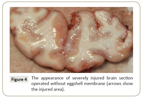
Thirty uncovered fresh cadaveric cow brain were used in this experimental feasibility study. In Group I (n=15), the number of minor injured brains was found to be 12 (80%). The appearance of minimally injured brain section operated with eggshell membrane is shown in Figure 3. In Group II (n=15), the number of minor injured brains was found to be 5 (33.33%). On the contrary, the number of moderately injured brains parenchyma in Group I cow brains was estimated to be 2 (13.33%). However, the number of moderately injured brains in Group II was found to be 9 (60%). The appearance of moderately injured brain section operated without eggshell membrane is shown in Figure 4. The number of severe injury was found to be 1 (6.67%) in Group II. The number of same injury was also found to be 1 (6.67%) in Group I. The evaluation of the sliced cow brain under the magnification of the operating microscope revealed that use of eggshell membrane protected the brain parenchyma compared with Group II specimens from the harmful effect of the microsurgical metallic instruments during the surgical intervention.
Discussion
Protecting the brain with its arterial and venous vascular structure and cranial nerves during microneurosurgical intervention is a critical and extremely important issue in the surgical practice of neurosurgery. Regional microneurosurgical neuro anatomy and microsurgical instruments should be well-known and recognized for a safe microneurosurgical intervention [1-4]. The use of these instruments with an appropriate microsurgical technique is crucial. It is imperative that surgical techniques should be repeated several times on appropriate models to successfully maintain and terminate microsurgical interventions including appropriately protecting of the neurovascular tissue [1-4]. There are some publications about the use of egg shell membrane in the human body. Chicken-egg shell membrane dressing significantly improves healing of cutaneous wounds in the early stages of wound healing. It is known that the egg shell membrane has an angiogenic capacity and antibacterial performance [5]. Egg shell membrane patching can be a good treatment choice to promote tympanic membrane healing in large traumatic tympanic membrane perforations [6].
Before performing a real operation on human beings, it is extremely necessary to have the capability of some metallic surgical devices to be used in the microneurosurgical intervention. Moreover, it is required for the person to develop his or her own abilities and create integrated personal surgical techniques for the appropriate protection of brain [1-4]. Vascular end to end, end to side, side-to-side anastomosis, aneurysm clipping and sylvian fissure dissection may be given as an example for microsurgical training models [3,4]. On the contrary, gaining detailed theoretical and practical microneurosurgical training on microsurgical models is not enough for brain protection. Advanced protection needs the use of some surgical materials during the surgical intervention. In the routine neurosurgical practice, cotton paddies, some elastic materials and limited use of brain retraction are all used for protecting of the brain.
In this experimental model, fresh cadaveric cow brains were used in the evaluating the efficacy of eggshell membrane for brain protection. An appropriate and successful model should have some similarities of the represented model. In contrast, another important issue is the easily obtainable and low-cost properties with the short and easy preparation of the model before use under the operating microscope without including any complicated steps. When taking into consideration the ethical issues, live models compromise some problematic limitations in experimental practice besides the aforementioned disadvantages. Some advantages are foreseen when evaluating the cow brain under the light of aforementioned parameters. Because the fresh cadaveric cow brain is not a living model, local ethical committee permission is not required. The fresh cadaveric cow brains were used in this study because of ethical convenience and similarities with the human brain.
Considering all these features together, the cow brain should be regarded as a suitable model in the experimental microneurosurgical brain protection. Few differences exist between the human and cow brains. The human brain is larger in size and shape when compared with the cow’s brain. Cow brains do not have as many gyri and sulci compared with human brains. The human brain of an adult weighs about 1200-1500 g and is 10 to 20 cm long. A cow’s brain is elongated in shape, whereas a human brain is rounded. However, some other differences exist in human and cow brains, but almost all mammalian brains are similar. Except some anatomical differences, the inter hemispheric sulcus and the arachnoid membrane of human and cow brains have the same characteristic features. In this experimental model, the similar microsurgical instruments were used during dissection, separation and distraction of the brain. Micro scissor, the tip of the micro aspirator and micro bayonet were used for the operation.
The operating site was on the left side in all-fresh cadaveric subjects. The left side of brain hemisphere was covered with the eggshell membrane. As the dissection progressed, the eggshell membrane was carefully pulled deep into the dissected and separated inter hemispheric sulcal space. The metallic brain component of the Leyla retractor was kept for 20 min. on the right hemisphere to retract the brain 2 cm lateral from the opposite hemisphere with standard chain retraction resistance. This was the final part of the experimental process. The presence of contusion, distortion and laceration were evaluated on the sliced brain materials using the operating microscope. The differences between protected and unprotected brain slices in terms of traumatic brain injury were quite clear. The protected brain hemispheres with egg shell membrane have less contusion, distortion and laceration injury compared with the unprotected brain hemispheres. Laceration and distortion are more common injuries in unprotected brain hemisphere.
Conclusion
This study showed that protecting the naked brain tissue with covering of eggshell membrane from the mechanical harmful effect of metallic microsurgical instruments is feasible. It is believed that this material might contribute to the practical micro neurosurgery in protecting brain tissue under the magnification of operating microscope.
References
- Cokluk C, Aydin K (2007) Maintaining microneurosurgical ability via staying active in microneurosurgery. Minim Invasive Neurosurg 50: 324-327.
- Altunrendke ME, Hamamcioglu MK, Hicdonmez T, Akcakaya MO, Birgili B, et al. (2014) Microsurgical training model for residents to approach to the orbit and the optic nerve in fresh cadaveric sheep cranium. J Neurosci Rural Pract 5: 151-154.
- Belykh E, Byvaltsev V (2014) Off-the-job microsurgical training on dry models: Siberian experience. World Neurosurg 82: 20-24.
- Spetzger U, von Schilling A, Brombach T, Winkler G (2011) Training models for vascular microneurosurgery. Acta Neurochir Suppl 112: 115-119.
- Guarderas F, Leavell Y, Sengupta T (2016) Assesment of chicken-egg membrane as a dressing for wound healing. Adv Skin Wound Care (United States) 29: 131-134.
- Jung JY, Yun HC, Kim TM (2017) Analysis of effect of egg-shell membrane patching for moderate-to-large traumatic tympanic membrane perforation. J Audiol Otol 21: 39-43.
Open Access Journals
- Aquaculture & Veterinary Science
- Chemistry & Chemical Sciences
- Clinical Sciences
- Engineering
- General Science
- Genetics & Molecular Biology
- Health Care & Nursing
- Immunology & Microbiology
- Materials Science
- Mathematics & Physics
- Medical Sciences
- Neurology & Psychiatry
- Oncology & Cancer Science
- Pharmaceutical Sciences


