Ventriculo-Peritoneal Shunt fracture : ( A case report)
Abdul Rahim H Zwayed1*, Sreenivas AV1, Amir M Shabana2, Yasser Abdul Rraziek3, Ban Farouk
Refaat4
Abdul Rahim H Zwayed1*, Sreenivas AV1, Amir M Shabana2, Yasser Abdul Rraziek3, Ban Farouk Refaat4
1Department of Neurosurgeon, Oman
2Consultant Anesthesia, Oman
3Consultant Radiologist, Oman
4Department of Pediatrician, Oman
*Corresponding author: Abdul Rahim H Zwayed, Department of Neurosurgeon, Oman, Tel: 96890122600; E-mail: zwayed62@yahoo.com
Received date: August 09, 2020; Accepted date: August 19, 2021; Published date: August 30, 2021
Citation: Zwayed ARH (2021) Ventriculo-Peritoneal Shunt fracture. Neurosurg Vol.5 No.3.
Abstract
Hydrocephalus (HC) is classically defined as dynamic imbalance between the production and absorption of cerebrospinal fluid (CSF) leading to enlarged ventricles. Ventriculo-peritoneal shunts (V.P.S.) are used to relieve hydrocephalus. Hydrocephalus can happen at any age, but it occurs more frequently among infants. There are a variety of congenital conditions that may cause cerebrospinal fluid to accumulate in the brain. These conditions include: aqueduct stenosis, encephaloceles, prematurity with germinal matrix hemorrhage. Complication have been reported, these are include: infections, shunt occlusion (common), disconnection and fracture (very rare).
Introduction
Hydrocephalus (HC) is classically defined as dynamic imbalance between the production and absorption of cerebrospinal fluid (CSF) leading to enlarged ventricles. Ventriculo-peritoneal shunts (V.P.S.) are used to relieve hydrocephalus.
Hydrocephalus can happen at any age, but it occurs more frequently among infants. There are a variety of congenital conditions that may cause cerebrospinal fluid to accumulate in the brain. These conditions include: aqueduct stenosis, encephaloceles, prematurity with germinal matrix hemorrhage.
Complication have been reported, these are include: infections, shunt occlusion (common), disconnection and fracture (very rare).
Here we report a case of VPS break in the chest region which is a very uncommon
We discuss the clinical picture, radiological findings and surgical procedure.
Key words: hydrocephalus, encephalocele, ventriculo-peritoneal shunt (VPS), break, and chest.
The case
This is a 6 years old girl, who was born of 40+ weeks as spontaneous vaginal delivery, with swelling in the occipital area.
There was a family history of brother with encephalocele operated on and did well.
On examination: (at birth): She was conscious, alert, active, flat anterior fontanel.
Head circumference of about =35.5 cm.
There was an occipital encephalocele of about 3x4 cm.
CT Head was done in the early days for more evaluation which report as:
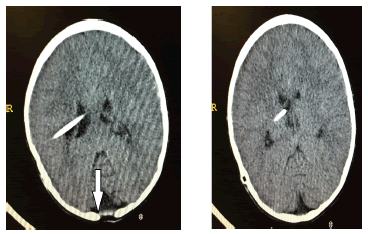
Occipital encephalocele with hydrocephalus.
So ventriculo-peritoneal shunt (VPS) done and repair of encephalocele
The girl stayed for 5 days then discharged in stable general condition
Follow up regularly the patient was stable, till at the age of 6 years, she presented with bouts of headache and swelling in the chest for about one week.
She gave history of vague athletic activity (as lifting her smaller brother) without history of direct trauma.
On examination: she was conscious, oriented, a febrile, no neurological deficits
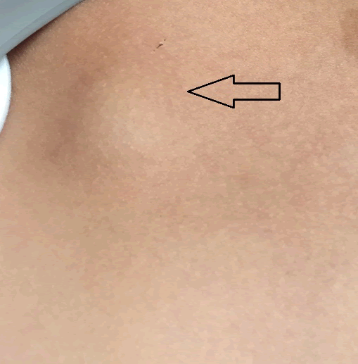
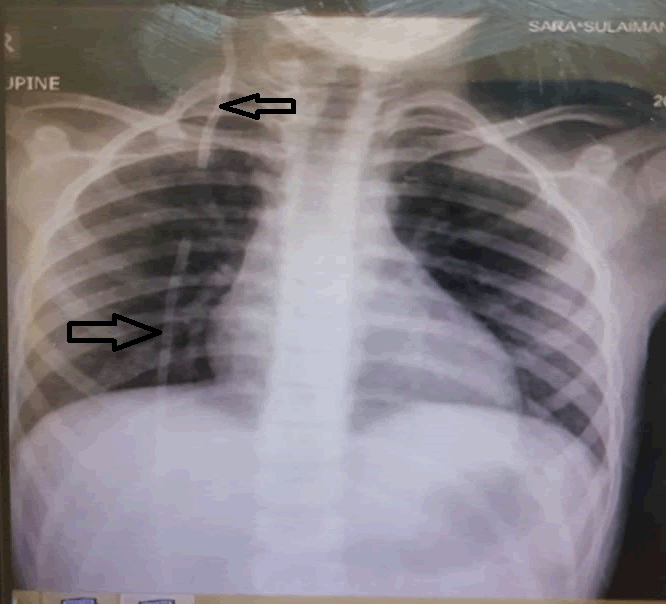
Local exam: There was an oval swelling of about 3 cm in diameter, in the upper chest with discontinuity of the shunt for about 10 cm
The device was compressible .There was no papilledema
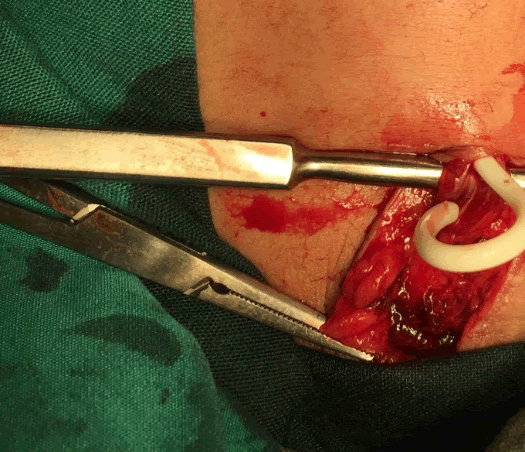
Revision of the lower end done with replacement of new peritoneal catheter. Patient stayed in the hospital and discharged after 3 days.
Follow up showed uneventful life.
Discussion
Hydrocephalus HC: is a condition that occurs when fluid builds up in the skull and causes the brain to swell, corresponds to an excessive accumulation of cerebrospinal fluid (CSF) within the central nervous system CNS, especially within the ventricles. Ventriculomegaly is the condition of enlarged cerebral ventricles. (3, 14)
Hydrocephalus can happen at any age, but it occurs more frequently among infants and adults 60 and over. The incidence of congenital HC is approximately 0.4–0.6/1,000 newborns with a slight downward trend. (14)
There are a variety of congenital conditions that may cause cerebrospinal fluid to accumulate in the brain. These conditions include:
Aqueduct stenosis (most common cause of congenital hydrocephalus), premature babies are at increased risk of intraventricular (germinal matrix) hemorrhage (is the most frequent cause of HC in infants.), encephaloceles, overproduction of fluid, or slow reabsorption of fluid. (14).
There are many underlying causes, but several are linked to autosomal and X-linked genetic disorders involving the CNS (11).
Hydrocephalus may produce an increase in intracranial pressure, which means high pressure within the skull. One of the ways to manage hydrocephalus is with a ventriculo-peritoneal shunt (VPS), which redirects the fluid away from the brain and to another area of the body that can more easily tolerate surplus fluid (10).
Ventriculoperitoneal shunt (VPS) is a medical device that relieves pressure on the brain caused by fluid accumulation (hydrocephalus) (2,14).
If the cause of the hydrocephalus is congenital (present from birth), or the result of a defect in the anatomy of the brain or spine, the VPS will be lifelong (2).
A VPS is a hollow tube with two openings, one on each end. One end of the tube is positioned underneath the skull, inside the ventricles, and the other end of the tube extends down through the body, with the opening positioned in the space that surrounds the abdominal region (the peritoneum). (5)
Ventriculo-peritoneal shunt (V.P.S.) which is used to relieve hydrocephalus have many complications rates which have been reported as high as 80% at any age. (13)
A patient with VPS need to maintain medical follow-up to avoid complications so that will recover as fully as possible.
These complications include, shunt obstruction, infection, and over drainage, malfunction, or blockage .Obstruction is the most common cause of Ventriculoperitoneal shunt (VPS) malfunction (8). Infection is the second most common cause of VPS malfunction, which is more common in children. . (7).
Pseudo cyst is a late complication of VPS, which may present as abdominal pain and a palpable mass.Bowel perforation is a rare complication of VPS that primarily occurs in premature infants and neonates. (12)
Subdural hematoma formation may occur with over-shunting in cases of low pressure hydrocephalus .Other complication like; migration (shunt must be pulled and have ability to move in the subcutaneous tissue, loose or improper connection may allow catheters to migrate. (9)
All previous mentioned complication are common, but complication as fracture (breakage of the catheter with separation of segments) is something rare
We report here a case of young girl with this complication after 6 years of the insertion of the VPS .Revision of the shunt done as removal of the peritoneal end (the 2 separate pieces) and replaced by new peritoneal catheter. She did well and follow up is uneventful life.
Disconnection and fracture: shunt disconnection/fracture comprises a rare cause of mechanical shunt malfunction (1)
Disconnection is defined as loss of continuity of shunt at normal connecting points between catheters, valves, and/or connectors
Fracture is actual breakage of the catheter with separation of segments
Associated factors for broken shunts are: growth spurts, aging, brittle or partially calcified shunt, multiple proximal revisions, local trauma to shunt, and athletic activity without history of direct trauma., post scoliosis correction ,shunt design (multiple shunt pieces have more risk of disconnecting. (11)
Most common location of breakage is in neck, followed by the scalp either proximal or distal to valve or connecting devices.
Patients with broken shunts may present with signs of increased intracranial pressure, pain, fluid collections along shunt tract and/or a palpable gap
Asymptomatic patients are usually diagnosed as an incidental radiological finding or during follow-up visits .Diagnosis of disconnection or fracture is confirmed on conventional radiographs (shunt series which show entire length of shunt from skull to abdomen).
CT scan can show increase in ventricular size or no change. (4, 6).
Treatment usually done by revision of this VPS.
References
- Blakeney William G, D`Amato, Charles MD (2015) Journal of Paediatric: Orthopedics â??Ventriculo peritoneal shunt fracture following application of Halo Gravity traction â?? A case report , EPT 35( 6): 52-54.
- CP Bondurant, DF Jimenez (1995) Epidemiology of cerebrospinal fluid shunting Pediatric. Neurosurgery 23 : 254-259.
- D Thompson, Hydrocephalus, shunts, AJ Moore, DW Newell (2005) Neurosurgery Principles and Practice, Specialist Surgery Series Neurosurgery. Springer 425-442 .
- Erica Roth(2017) what is a Ventriculoperitoneal shunt
- JJ Stone, CT Walker (2013) Revision rate of pediatric ventriculo-peritoneal shunts after 15â?¯years, J. Neurosurg. Pediatr 11: 15-19.
- JM Drake, JR Kestle, Piatt JJ (1998) Randomized trial of cerebrospinal fluid shunt valve design in pediatric hydrocephalus. Neurosurgery 43: 294-305.
- J Kestle, J Drake, F Boop (2000) Long-term follow-up data from the shunt design trial. Pediatr Neurosurg 33: 230-236.
- JW Cozzens, JP Chandler (1997) Increased risk of distal ventriculo-peritoneal shunt obstruction associated with slit valves or distal slits in the peritoneal catheter. J Neurosurg 87: 682-686 .
- MJ McGirt, DW Buck II, J Weingart (2007) Adjustable vs. set-pressure valves decrease the risk of proximal shunt obstruction in the treatment of pediatric hydrocephalus. Childs Nerve Syst 23: 289-295.
- MJ McGirt, J Leveque, JS Hopkins, HE Fuchs (2002) Cerebrospinal fluid shunt survival and etiology of failures: a seven year institutional experience. Pediatric Neurosurgery 36: 248-255.
- M. Verhagen, C.T. Schrander-Stumpel, et al.Congenital hydrocephalus in clinical practice: a genetic diagnostic approach, European Journal of Medical Genetics, 54 (2011), pp. e542-e547
- Rainov, Heidecke V. Schobess: Abdominal CSF pseudocysts in patients with ventriculo peritoneal shunts. Report of fourteen cases and review of the literature ,Acta Neurochir., 27 (1â??2) (1994), pp.73-78.
- Reddy GK, Bollam P, and Caldito, G.: Long term outcomes of ventriculo-peritoneal shunt surgery in patients with hydrocephalus. World Neurosurg. 01.96: 404â??410.
- Thakkar R., Lawrence M. Shuer, , Hydrocephalus. American Association of Neurological Surgeons. http://www.aans.org/en/Patients/Neurosurgical-Conditions-and-Treatments/Hydrocephalus. Accessed June 1, 2017.
Open Access Journals
- Aquaculture & Veterinary Science
- Chemistry & Chemical Sciences
- Clinical Sciences
- Engineering
- General Science
- Genetics & Molecular Biology
- Health Care & Nursing
- Immunology & Microbiology
- Materials Science
- Mathematics & Physics
- Medical Sciences
- Neurology & Psychiatry
- Oncology & Cancer Science
- Pharmaceutical Sciences
